
Clinical case presented by Dr. Roccuzzo Italy
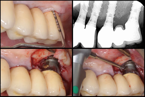
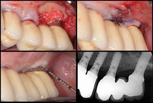
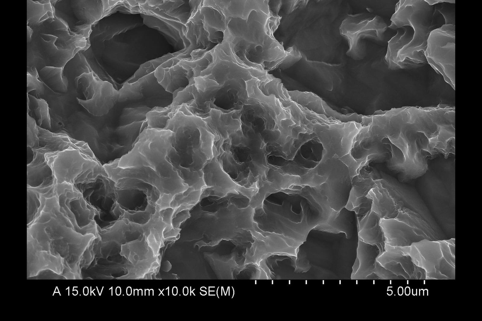
Implant surface under SEM, before and after
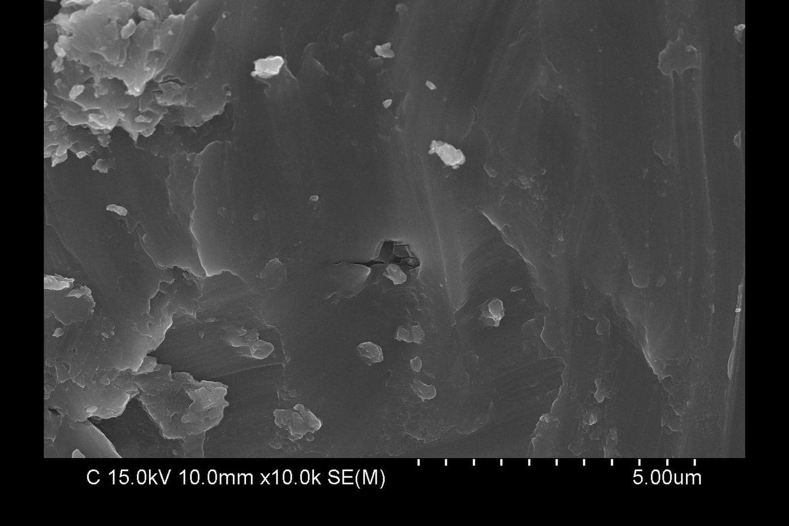
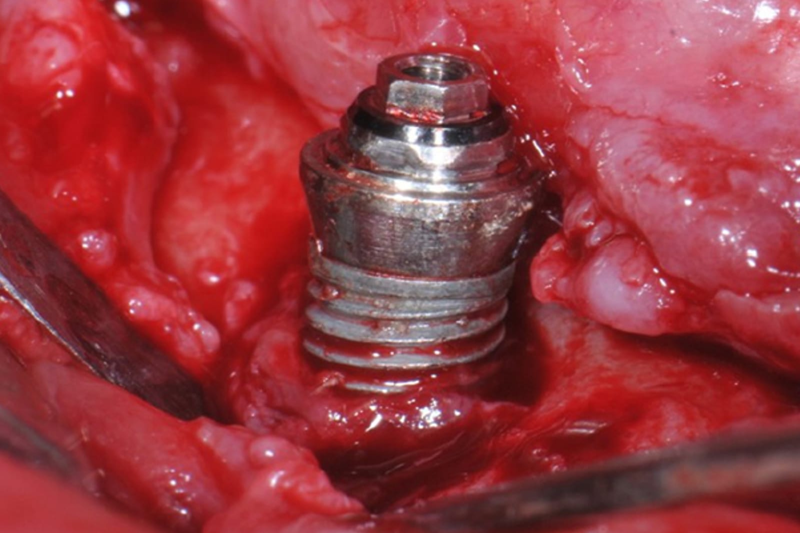
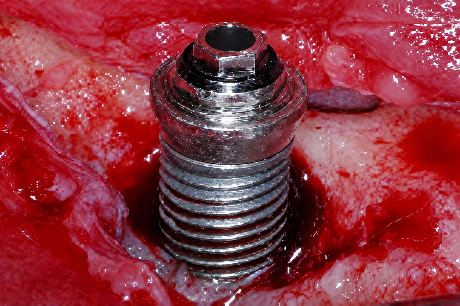
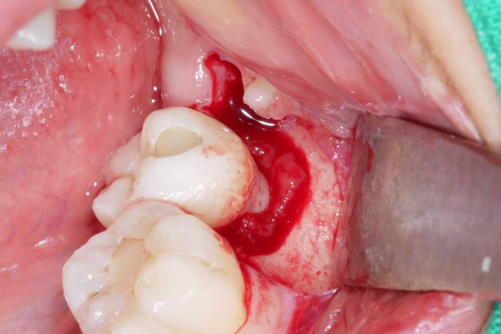
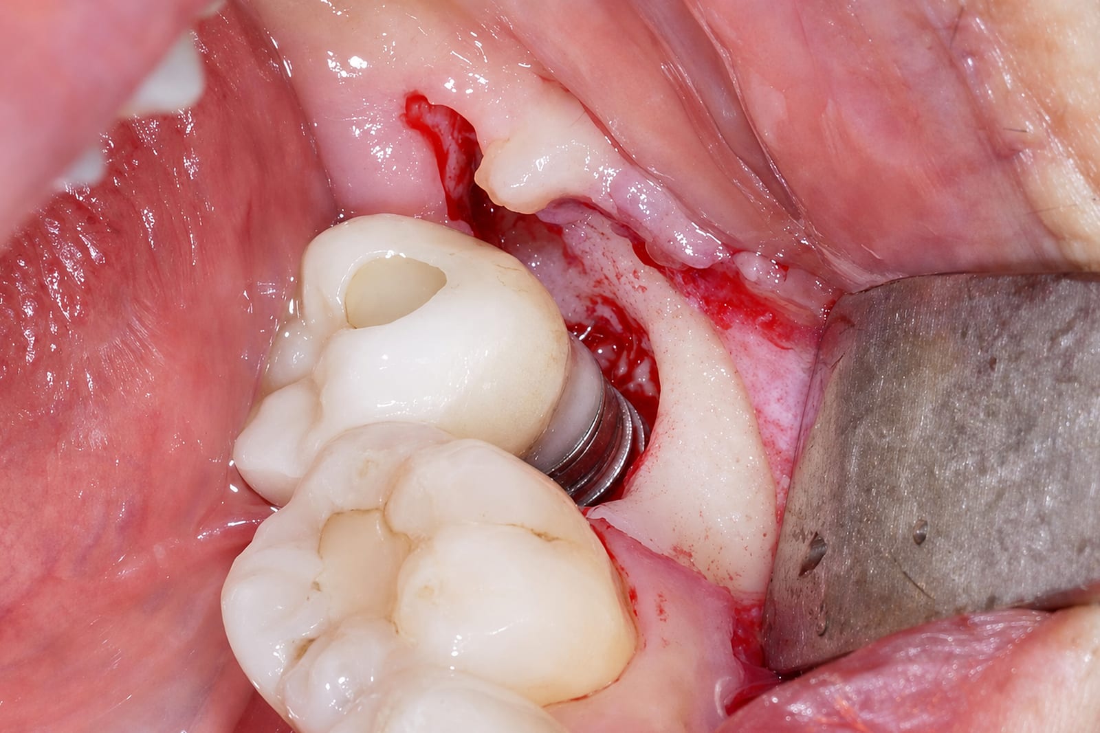
Surface after debridement intra-operative
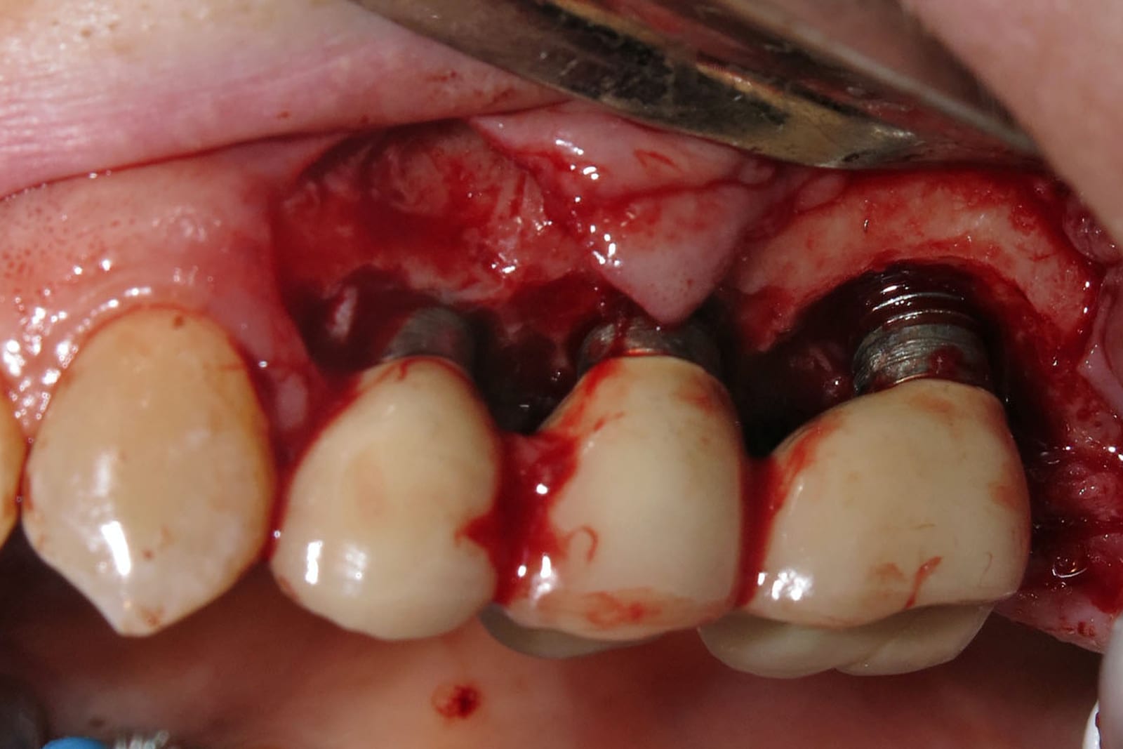
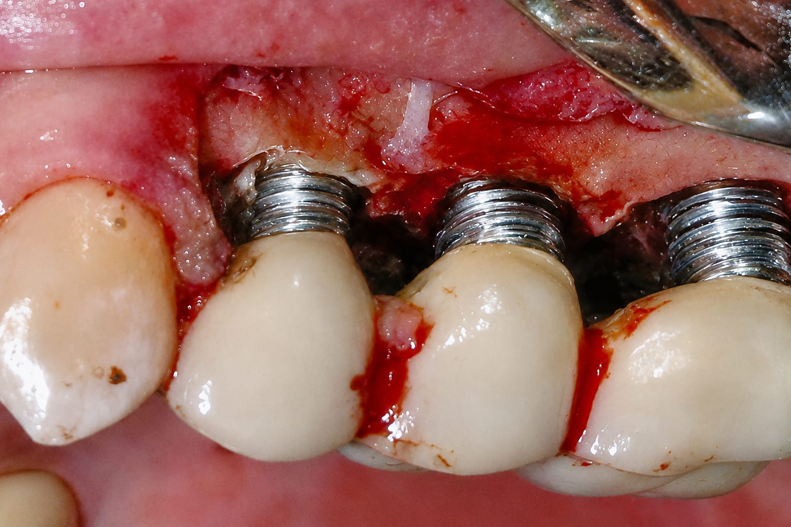
The exposed threads before treatment, and the same implant once the contaminated layer has been removed with the NiTi Brush — the surface returns to a bright, smooth† finish.
Clinical case presented by Prof. Baek-Soo Lee
Department of Oral and Maxillofacial Surgery, Kyung-Hee University, Seoul, Korea.
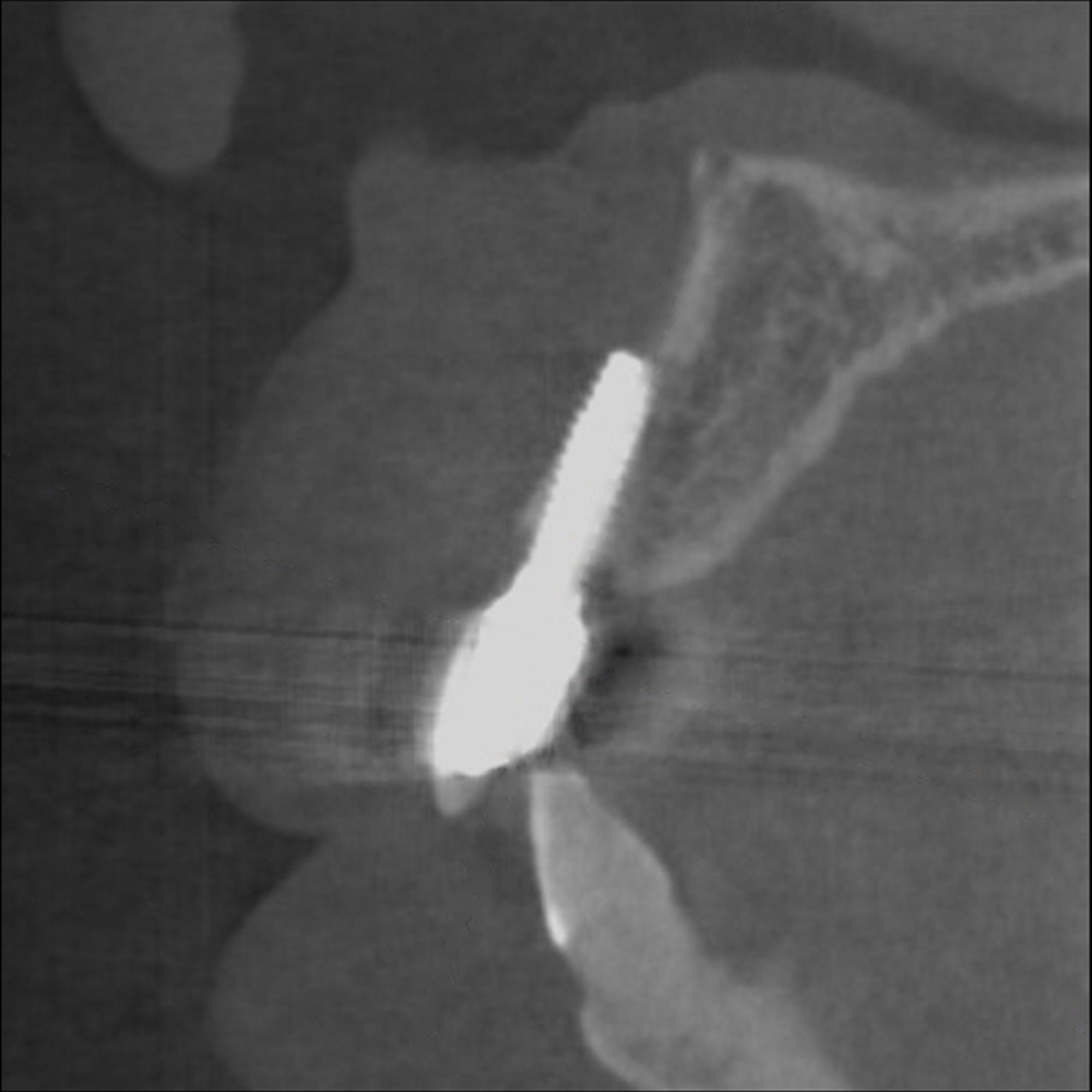
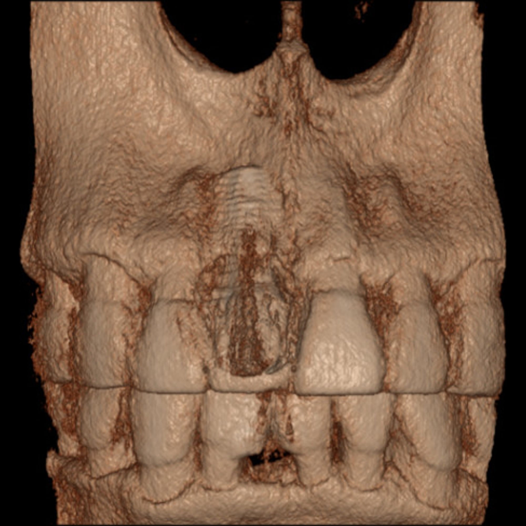
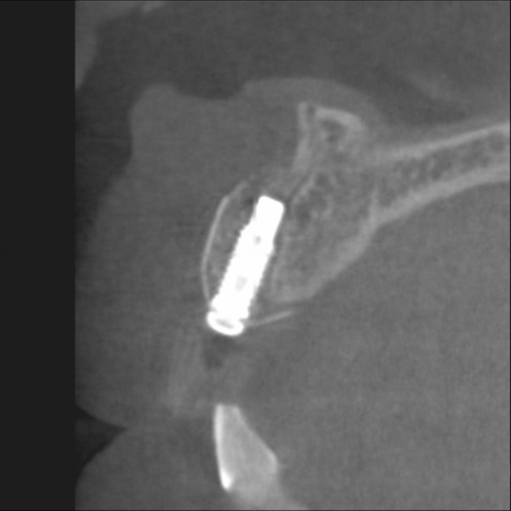
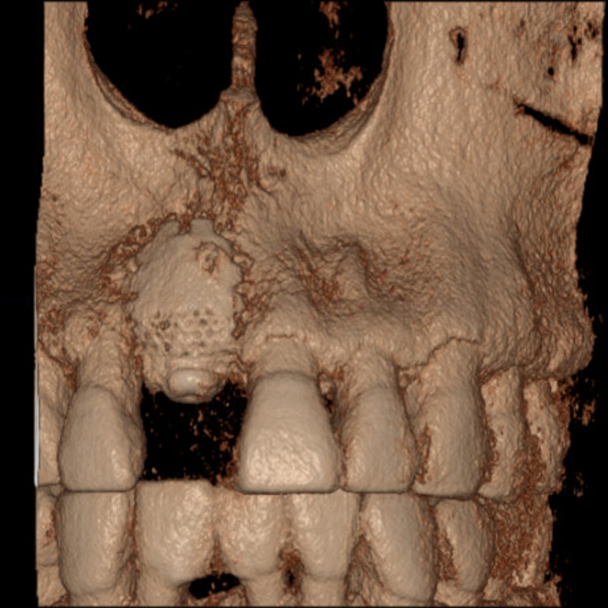
Before and after, animated 45 seconds, no sound
The CBCT sections and 3D reconstructions above, compared side by side: bone loss around the implant before treatment, and new bone formation after debridement with the NiTi Brush and guided bone regeneration. Plays on this site directly; press play to start.
Clinical case 01
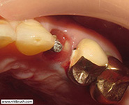
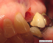
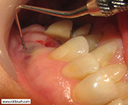
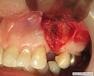
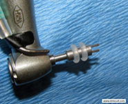
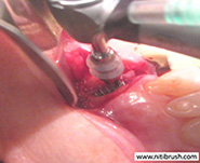
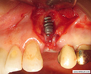
GBR sequence
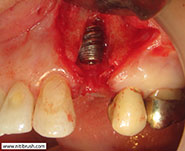
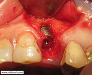
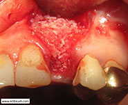
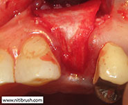
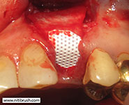
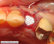
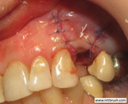
Presented by Dr. Frederick Li Vancouver, Canada

Clinical case 02
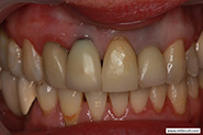
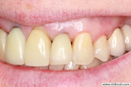
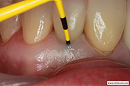
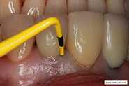
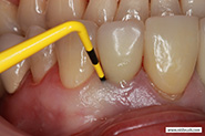
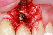
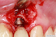
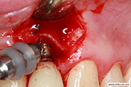
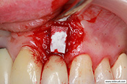
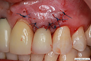
#periimplantitis #niti brush #Titanium brush #Titanium brush for peri-implantitis #niti brush for peri-implantitis #R brush #I brush #Rotating titanium brush #peri brush
Note on “smooth”
† Smooth refers to moderately rough implant surfaces (e.g. SLA, RBM). In laboratory studies with the NiTi Brush, surface micro-roughness was reduced by removing protrusions (Kim et al. 2016) and the sharp-edged microstructure was smoothed, brushing continuing until the surface looked uniformly smooth and shiny, as in clinical use (Brunello et al. 2025). A clinical review describes polishing the exposed rough implant surface to a smooth surface with the NiTi Brush (Hiroyasu 2018). The shine seen chairside is the visible sign of this change (HANS Korea performance test).