

The clinical problem and why it is growing
Implant dentistry has been placing implants at scale for three decades. The population of implants now old enough to fail is larger every year — and peri-implantitis is the reason most of them do.
When removing the implant is not a realistic option
A single, standalone implant in a healthy patient can usually be explanted and replaced. That option narrows sharply in the cases that actually present in practice — and it is rarely the number of failing implants that decides the difficulty.
The practical alternative is to save what is already there
In these cases the realistic path is not removal and replacement — it is to decontaminate the exposed surface and keep the implant in function. That is the problem the NiTi Brush was designed to solve.
It is also the principle the company was founded on: manage and treat the implant like a natural tooth — do not remove the implant.
What peri-implantitis is
Peri-implantitis is an inflammatory condition of the tissues around an osseointegrated dental implant, characterised by inflammation of the peri-implant mucosa together with progressive loss of the supporting bone.3
It is distinguished from peri-implant mucositis, in which the inflammation is confined to the soft tissue and no supporting bone has yet been lost. Peri-implant mucositis is considered the precursor of peri-implantitis, in the same way that gingivitis precedes periodontitis.
The condition is also written periimplantitis or peri implantitis, and is sometimes referred to more broadly as peri-implant disease or implantitis.
Clinical signs
- Bleeding and/or suppuration on gentle probing
- Increased probing depth compared with earlier examinations
- Radiographic bone loss beyond initial remodelling
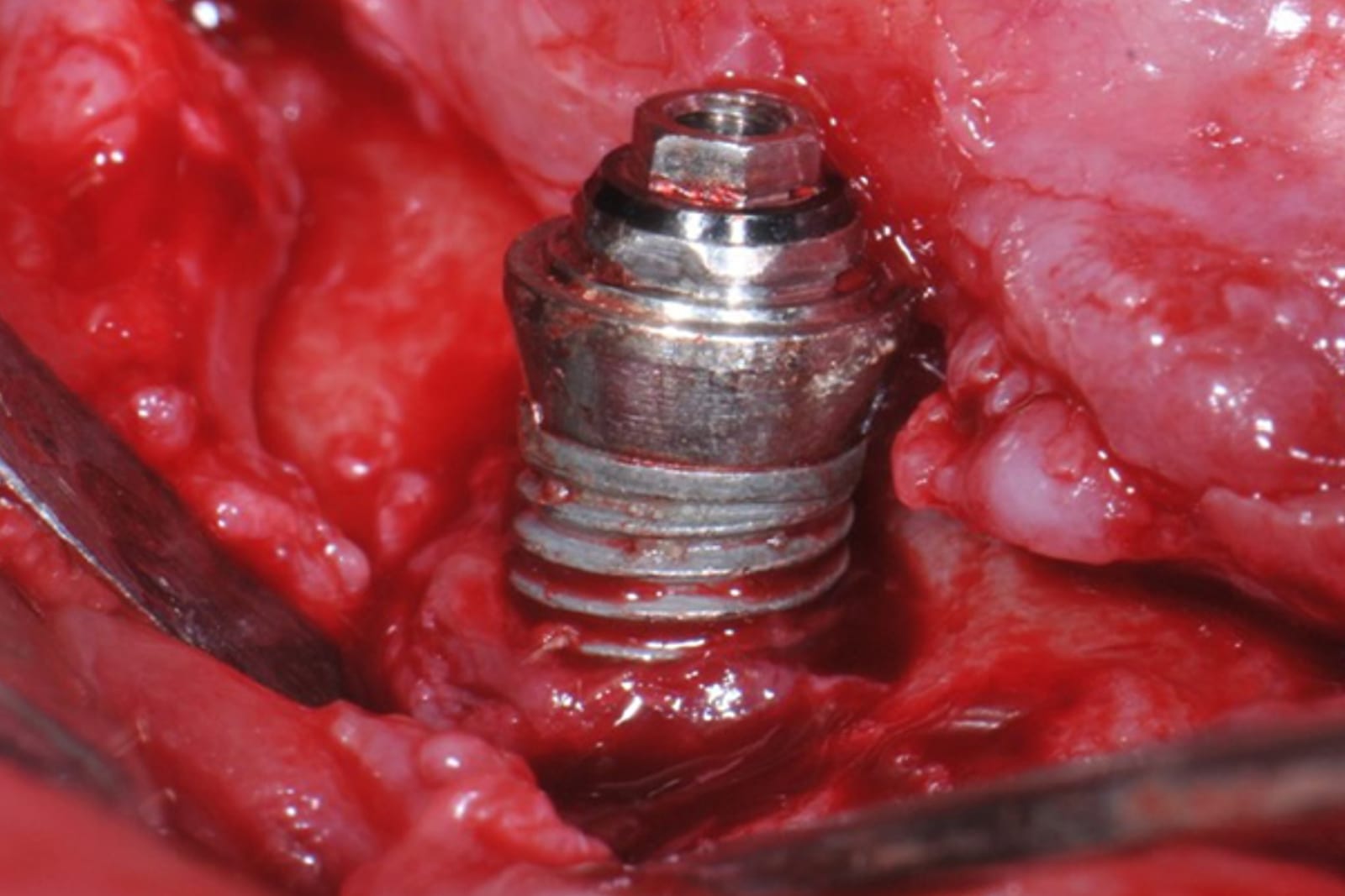
- Exposed, contaminated implant threads once a flap is raised
Why the implant surface is the problem
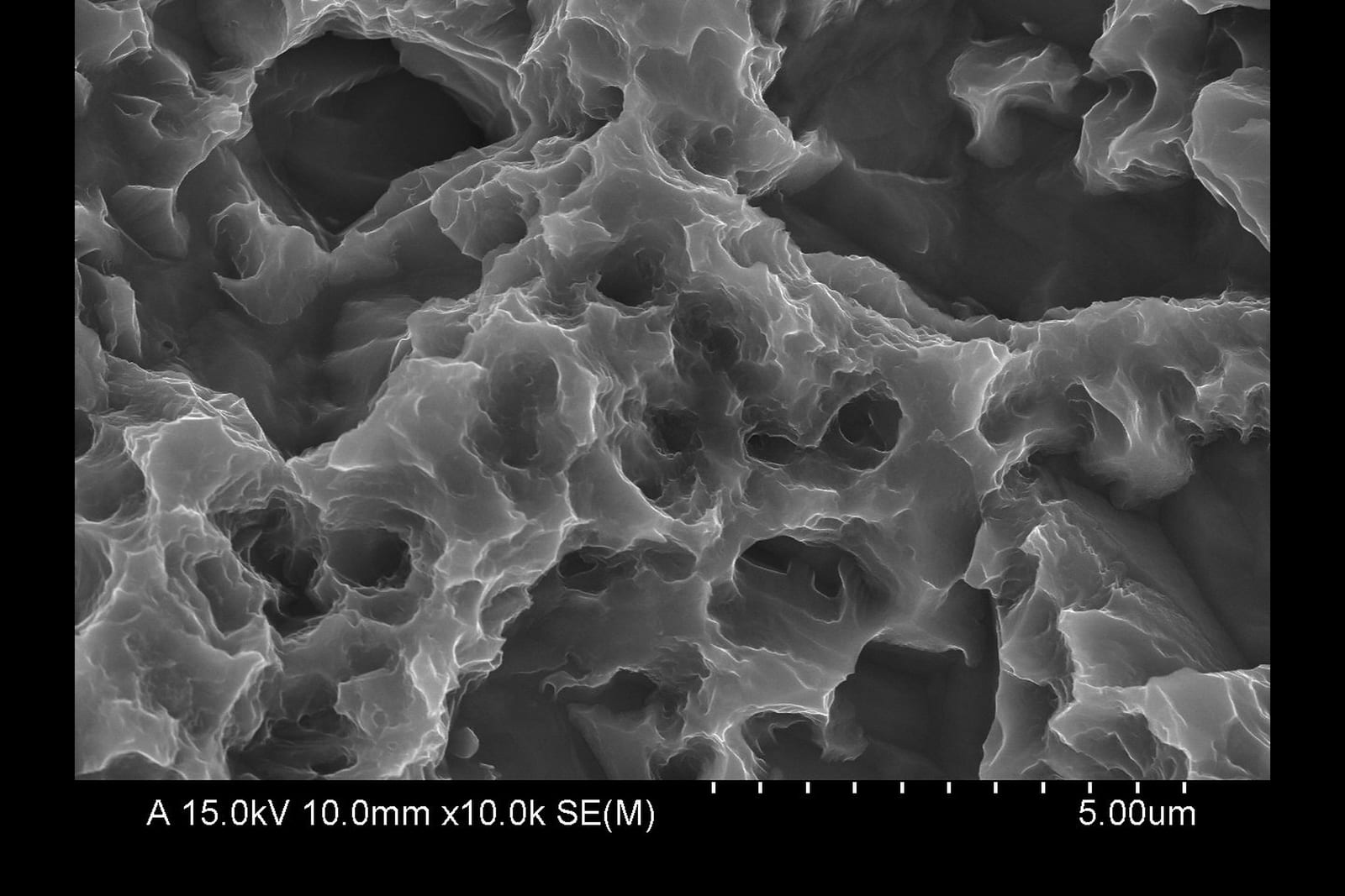
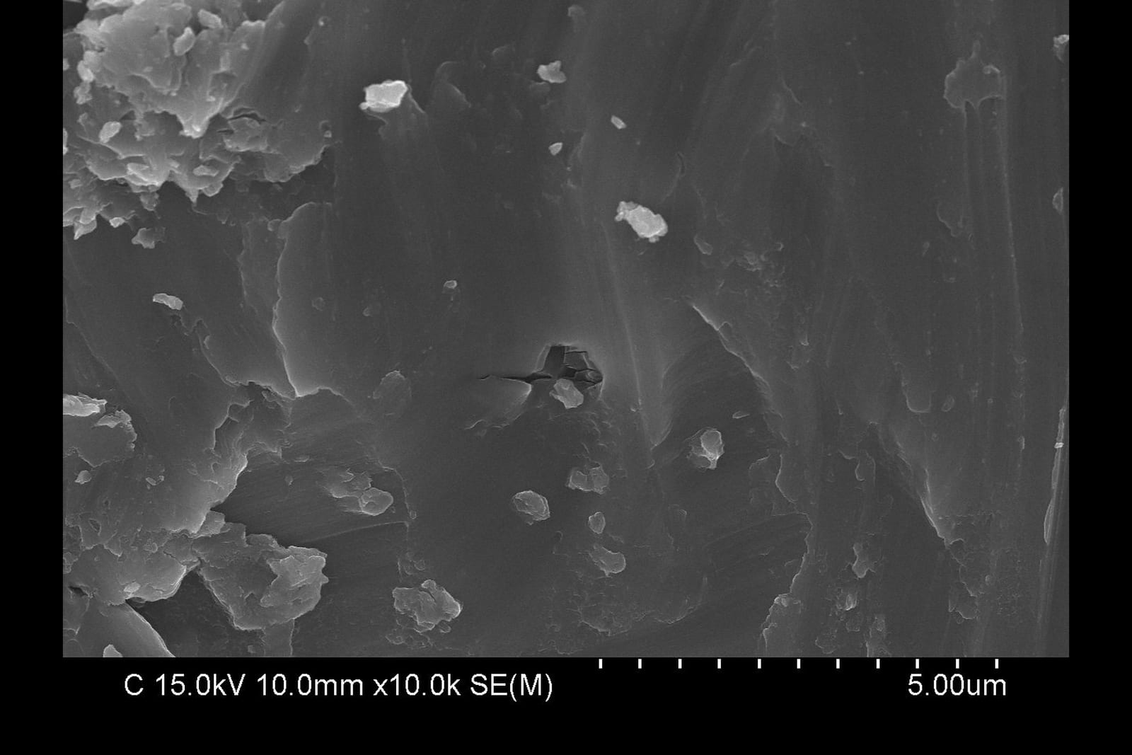
Modern dental implants are deliberately given a roughened surface, because roughness improves osseointegration. The same roughness works against the clinician once the surface becomes exposed: bacterial biofilm anchors into the micro-topography and into the thread profile, where neither instrumentation designed for smooth root surfaces nor irrigation alone can reliably reach it.
This is why the decontamination step, rather than the regenerative material, is often what decides the result of surgical therapy. A graft or membrane placed against a surface that is still contaminated is placed against a surface that will keep the inflammation going.
Approaches to implant surface decontamination
The literature describes mechanical, chemical and physical approaches, most often used in combination. They are not mutually exclusive; the mechanical step is what physically removes the deposit, and the others support it.
| Approach | Examples | Role |
|---|---|---|
| Mechanical treatment | Mechanical debridement and polishing of the surface using the NiTi Brush for peri-implantitis | Physically removes biofilm, calculus and granulation tissue from the implant surface, and removes the roughened surface itself — together with mechanical removal of granulation tissue from the implant surface and the surrounding tissue |
| Chemical | Chemical treatment after brushing | Refer to the references and literature |
| Reconstructive | GBR — guided bone regeneration | Refer to the references and literature |
What the NiTi Brush is for
The first purpose of the NiTi Brush for peri-implantitis is mechanical detoxification, mechanical decontamination and mechanical cleaning of the implant surface. Osseous defects of plaque, calculus and any other foreign body material resulting from peri-implantitis are detoxified, decontaminated and cleaned mechanically by the brush.
The second purpose is the mechanical removal or debridement of granulation tissue — both from the implant surface and from the tissue surrounding the implant.
Newer decontamination methods are tested on top of mechanical debridement
When a chemical adjunct or a reconstructive material is evaluated, the trial design places it alongside mechanical instrumentation, not instead of it. In the randomised trials that established the current protocols, the mechanical step was performed with a NiTi brush.4
In one such trial the protocol specified surface decontamination “by means of NiTi brushes for about 2–3 min at 600 rpm” before the adjunct was applied.4
Peer-reviewed studies on these approaches — including randomised controlled trials on reconstructive therapy and on the significance of barrier membranes — are listed on the Clinical evidence page.
Mechanical debridement with a nickel-titanium brush
A rotary brush addresses a specific difficulty: the thread profile. A bristle that is stiff enough to remove the deposit but flexible enough to follow the thread can reach into the undercut geometry that a rigid instrument passes over.
Nickel-titanium is used for this reason. With a modulus of elasticity of roughly 28–41 — against 115 for titanium and 210 for stainless steel — Ni-Ti is about three times more flexible than titanium, while its Vickers hardness of 303–362 is comparable to titanium's 200–340. The bristle bends into the thread rather than cutting across it. Stainless steel, at 600–610 Vickers and far stiffer, is not a suitable bristle material for a titanium implant surface.
What the operator sees
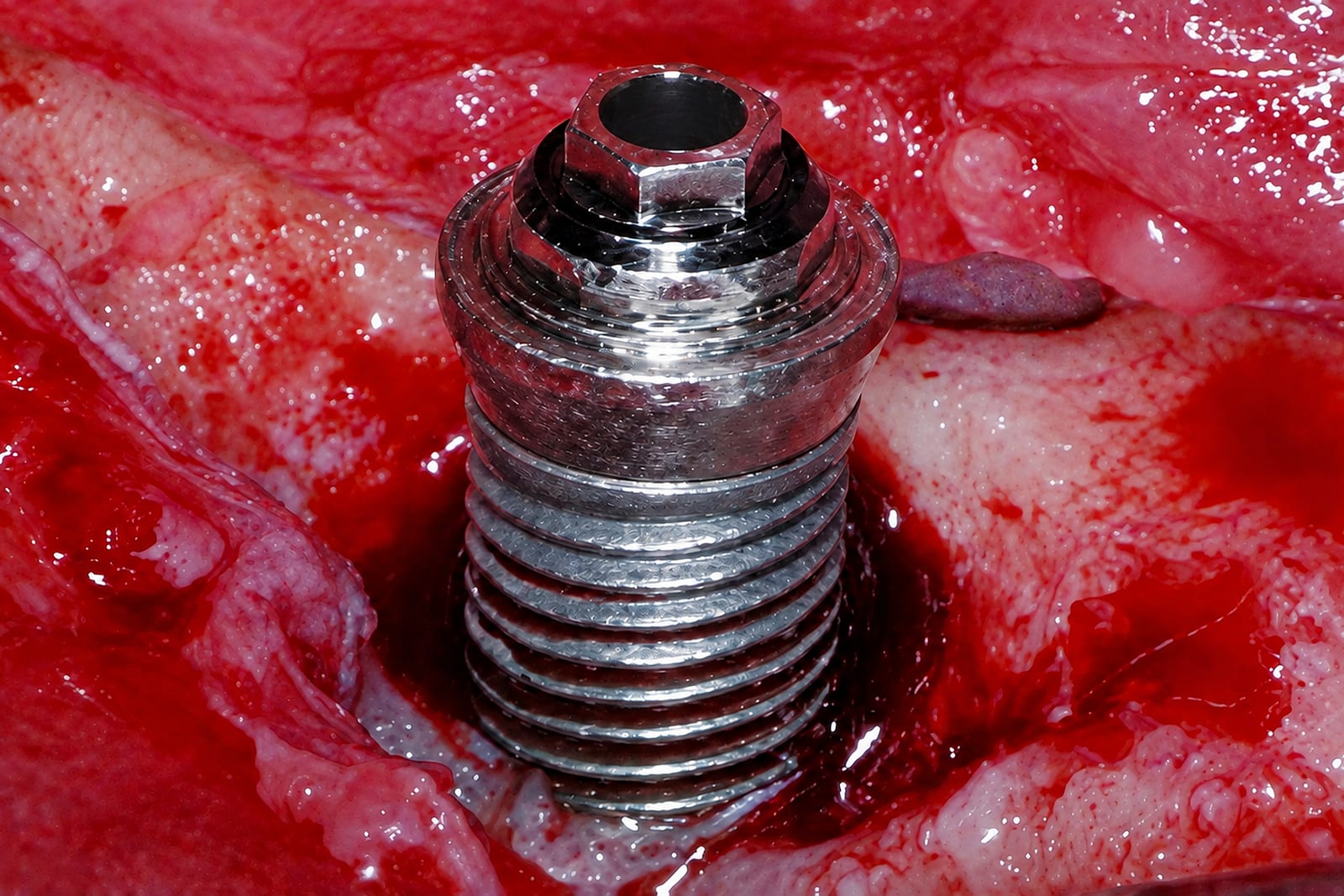
During brushing the shade of the implant surface changes: the dull, contaminated appearance gives way to a bright, machined finish. This visible change is the chairside endpoint of the debridement step.
The NiTi Brush protocol example of protocol
-
Diagnosis
Diagnose peri-implantitis and measure the depth of the pocket.
-
Select the brush
NANO for 1–2 threads exposed · POCKET for more than 2 threads · POCKET SHORT for molars.
-
Sterilise
Supplied non-sterile. Autoclave in a sterile pouch at 134 °C for 15 minutes.
-
Rotate — debridement & decontamination
Implant motor, 600–1,200 rpm with extensive external saline. Turn the rough surface into a machined smooth surface, treating the implant surface and the surrounding tissue at the same time. Premium-brand implants take about 30–40 seconds; other brands 1–3 minutes.
-
Chemical treatment
Tetracycline 250 mg in 2.5 cc warm saline, applied by syringe to the brushed surface and tissue. Wait 3 minutes, then wash away with saline.
-
GBR
Guided bone regeneration follows the chemical step — refer to the literature.
Use an implant motor. The brush is driven through a 20:1 reduction handpiece at 600–1,200 rpm with continuous external sterile saline. It is not intended for a high-speed turbine or any drive that cannot hold this speed range.
Supplied non-sterile — autoclave at 134 °C for 15 minutes before every use. Single use only, do not reuse. This summary does not replace the Instructions for Use. Always read the IFU applicable to your market before treatment.
Choosing the brush for the defect
Four geometries cover the range of access encountered in practice — from a single exposed thread through to deep pocket involvement.
| Model | Bristle head | Thread exposure | Stage |
|---|---|---|---|
| NANO | Pointed, bound | One thread open | Initial |
| Open | 2–3 threads open | Established | |
| POCKET SHORT | Open, short shaft | 2–3 threads open | Narrow posterior / limited opening |
| OMEGA | Wide, splayed | Deep pocket | Advanced |
References
- Derks J, Tomasi C. Peri-implant health and disease. A systematic review of current epidemiology. Journal of Clinical Periodontology 2015;42(Suppl 16):S158–S171. DOI: 10.1111/jcpe.12334
- Montero E, Roccuzzo A, Molina A, Monje A, Herrera D, Roccuzzo M. Minimal invasiveness in the reconstructive treatment of peri-implantitis defects. Periodontology 2000 2022. DOI: 10.1111/prd.12460
- Berglundh T, Armitage G, Araujo MG, et al. Peri-implant diseases and conditions: consensus report of workgroup 4 of the 2017 World Workshop on the Classification of Periodontal and Peri-Implant Diseases and Conditions. Journal of Clinical Periodontology 2018;45(Suppl 20):S286–S291. DOI: 10.1111/jcpe.12957
- Monje A, Pons R, Vilarrasa J, Nart J, Wang H-L. Significance of barrier membrane on the reconstructive therapy of peri-implantitis: a randomised controlled trial. Journal of Periodontology 2023. DOI: 10.1002/JPER.22-0511
HANS Korea is not the publisher of these works and has no commercial relationship with them. See the legal notice.
peri-implantitis · periimplantitis · peri implantitis · peri-implant disease · peri-implant mucositis · implant surface decontamination · implant surface debridement · mechanical debridement · titanium brush · NiTi brush · nickel-titanium brush · rotating titanium brush · implantoplasty · guided bone regeneration